Breast cancer remains one of the most diagnosed cancers worldwide, yet survival rates have improved dramatically over the past three decades. This shift stems largely from advances in screening technology and a better understanding of when and how to detect abnormalities before they progress. For women navigating their health options, knowing what screening involves and when to seek diagnostic imaging can be the difference between catching cancer at stage one versus stage three.
The Role of Imaging in Early Detection
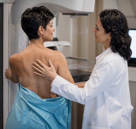
Screening mammography forms the foundation of breast cancer detection programs. Standard 2D mammograms have been in use for decades, but the technology has evolved considerably. Three-dimensional mammography, also called tomosynthesis, creates multiple thin-slice images of breast tissue rather than a single flat image. This layered approach reduces the problem of overlapping tissue that can obscure small masses or create false alarms.
For women with dense breast tissue, mammography alone may not provide sufficient detail. Dense tissue appears white on mammograms, as do tumors, making it harder to distinguish abnormalities. In these cases, supplemental screening with breast MRI or ultrasound becomes valuable. MRI uses magnetic fields and radio waves to produce detailed images and is particularly effective for high-risk women, including those with BRCA gene mutations or a strong family history of breast cancer.
When Screening Leads to Diagnosis
Not every abnormal screening result indicates cancer. Radiologists use the BI-RADS classification system to categorize findings, with scores ranging from 0 (incomplete assessment) to 6 (known biopsy-proven malignancy). A BI-RADS 4 or 5 finding typically requires a biopsy to determine whether cells are cancerous.
Image-guided biopsy has replaced many surgical biopsies as the diagnostic standard. During this procedure, imaging technology directs a needle to the precise location of the abnormality. Core needle biopsy removes small cylinders of tissue, while vacuum-assisted biopsy can remove larger samples. Both techniques are performed under local anesthesia and leave minimal scarring compared to surgical alternatives. The tissue sample then goes to pathology for analysis, which determines whether cancer is present and, if so, what type and grade.
Understanding Biopsy Results
Pathology reports contain critical information that guides treatment decisions. The report identifies whether the cancer is invasive or in situ, meaning it has remained within the milk ducts or lobules. It also tests for hormone receptors (estrogen and progesterone) and HER2 protein status. These markers determine whether the cancer will respond to hormone therapy or targeted treatments like trastuzumab.
Comprehensive Treatment Approaches
Once diagnosis is confirmed, treatment planning involves a multidisciplinary team including surgical oncologists, medical oncologists, radiation oncologists, and reconstructive surgeons. The goal is to eliminate cancer while preserving quality of life and, when possible, breast appearance.
Surgical options have expanded beyond traditional mastectomy. Lumpectomy, or breast-conserving surgery, removes the tumor and a margin of surrounding tissue while leaving most of the breast intact. This approach typically requires follow-up radiation therapy. For women requiring mastectomy, robotic-assisted techniques offer improved precision and potentially faster recovery compared to conventional surgery. The choice between lumpectomy and mastectomy depends on tumor size, location, genetic factors, and patient preference.
Medical oncology has seen remarkable advances in targeted therapies and immunotherapy. Unlike traditional chemotherapy, which affects all rapidly dividing cells, targeted therapies attack specific molecular pathways that cancer cells use to grow. For HER2-positive cancers, monoclonal antibodies block growth signals. For hormone receptor-positive cancers, endocrine therapies prevent estrogen from fueling tumor growth. However, recent research has highlighted that hormonal therapies may increase cardiovascular risk in some patients, underscoring the importance of monitoring overall health during treatment.
Accessing Specialized Breast Health Services
Women in the Gulf region seeking comprehensive care have access to specialized programs that integrate screening, diagnosis, and treatment under one coordinated system. For those in the UAE capital, Cleveland Clinic Abu Dhabi’s Breast Health & Cancer Program offers services ranging from advanced imaging technologies like 3D mammography and breast MRI to genetic testing for hereditary cancer risk. Their approach includes medical oncology treatments such as targeted therapies and immunotherapy, surgical options including robotic mastectomy, and breast reconstruction using both implant-based and autologous tissue techniques like the DIEP flap procedure. Women can consult with a breast surgeon in abu dhabi who works as part of a multidisciplinary team to develop individualized treatment plans based on each patient’s specific diagnosis and circumstances.
Reconstruction and Recovery
For women undergoing mastectomy, breast reconstruction can be performed immediately during the same surgery or delayed until after completion of cancer treatment. Implant-based reconstruction uses saline or silicone implants to recreate breast shape. Autologous reconstruction transfers tissue from another part of the body, most commonly the abdomen in a procedure called DIEP flap reconstruction. This technique preserves abdominal muscles while using skin, fat, and blood vessels to construct a new breast.
The choice between reconstruction methods involves trade-offs. Implants require less extensive surgery and shorter initial recovery but may need replacement after 10 to 15 years. Autologous reconstruction involves longer surgery and recovery time but produces results that age naturally with the body and typically do not require replacement. Some women choose no reconstruction, opting instead for external prostheses or embracing their post-surgery appearance.
Managing Long-Term Effects
Lymphedema, or swelling caused by lymph fluid buildup, can develop after lymph node removal or radiation therapy. Prevention strategies include avoiding blood pressure measurements and injections in the affected arm, maintaining a healthy weight, and performing prescribed exercises. Early intervention through specialized lymphedema therapy can prevent progression and reduce symptoms.
Radiation therapy, while highly effective at reducing local recurrence, can cause fatigue, skin changes, and in some cases, long-term effects on breast appearance or shoulder mobility. Modern techniques like intensity-modulated radiation therapy (IMRT) and proton therapy aim to minimize exposure to surrounding healthy tissue, particularly the heart and lungs. Women who have undergone radiation therapy should discuss ongoing monitoring for potential late effects with their oncology team.
Genetic testing plays an increasingly important role in breast cancer care. Women with BRCA1 or BRCA2 mutations face significantly elevated lifetime breast cancer risk and may benefit from enhanced surveillance or risk-reducing surgery. Testing also informs treatment decisions, as some targeted therapies work specifically in BRCA-mutated cancers. Family members can use genetic information to make informed decisions about their own screening and prevention strategies, creating a framework for better outcomes across generations.
The Future of Breast Cancer Care
Advances in precision medicine continue to refine treatment selection. Genomic tests like Oncotype DX analyze tumor genes to predict recurrence risk and whether chemotherapy will provide benefit beyond hormone therapy alone. For women with early-stage, hormone receptor-positive cancer and low recurrence scores, this can mean avoiding chemotherapy and its side effects without compromising outcomes.
Artificial intelligence is beginning to assist radiologists in detecting subtle abnormalities on mammograms and predicting which lesions are most likely to be cancerous. These tools do not replace human expertise but rather augment it, potentially reducing both missed cancers and unnecessary biopsies. As the technology matures, it may help address radiologist shortages and improve consistency in screening interpretation.
The trajectory of breast cancer care reflects a shift from one-size-fits-all protocols to individualized approaches based on tumor biology, genetic factors, and patient preferences. Screening remains the cornerstone of early detection, but what happens after an abnormal result now involves sophisticated imaging, precise diagnostic techniques, and an expanding array of treatment options that balance cancer control with quality of life.